
The Ritchey Chrétien telescope, a reflecting telescope in the Cassegrain system, reduces spherical aberration and coma for clearer viewing.
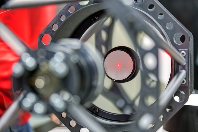
The Ritchey Chrétien telescope, a reflecting telescope in the Cassegrain system, reduces spherical aberration and coma for clearer viewing.
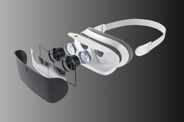
The triplet apochromat lens in VR headsets redefines immersion with minimal chromatic aberration, offering unparalleled clarity and realism.

Explore how the 50X long working distance objective maintains superior and impressive magnification performance, catering to your specific and demanding applications.
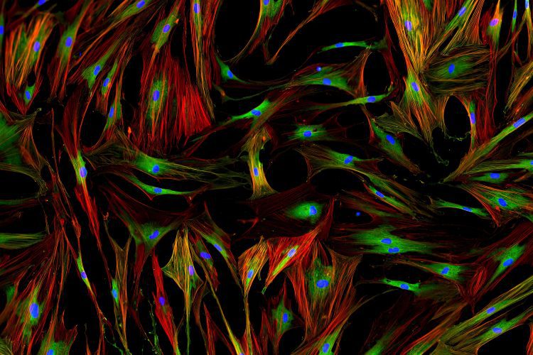
Oblique Illumination, AI algorithms, and big data analytics enhance Live Cell Imaging by improving cellular visualization and analysis.

Total internal reflection (TIR) is a phenomenon in physics where light rays travel from a more optically denser medium to a less optically denser medium.

Light and optics study how light interacts, including Total Internal Reflection and the critical Angle of Refraction in denser mediums.

Flow cytometry relies on optimizing SNR to enhance the clarity of signals from cells using lasers and flow cytometer optics.

The guide explains spherical mirrors, the mirror equation, radius of curvature, and contrasts convex and concave mirrors.
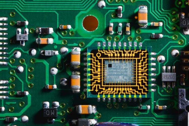
PICs, or optical ICs, use photons to power highly sensitive biosensors and revolutionize medical diagnostics.
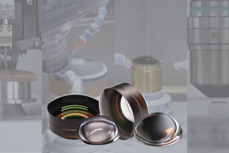
One of the most important features of aspheric lenses is their ability to correct for spherical aberration. Spherical aberration is found in all spherical lenses, such as plano-convex or double-convex lens shapes.