Diffuse Optical Tomography (DOT) is a non-invasive imaging technique using near-infrared light to visualize optical properties of soft tissue, such as brain or breast tissue.
By analyzing light absorption and scattering due to chromophores like hemoglobin, DOT enables real-time imaging of tissue oxygenation and composition.
Techniques include time, frequency, and steady-state domain systems.
Though its spatial resolution is lower than MRI, DOT is portable, cost-effective, and valuable in neurology, oncology, and surgery.
Avantier offers custom optics tailored for DOT applications.
Diffuse Optical Tomography (DOT) is a medical imaging technique that uses NIR light to measure the optical properties of translucent or semi-translucent biological tissue. That includes most soft tissue— breast and brain tissue, for instance, are good candidates for DOT.
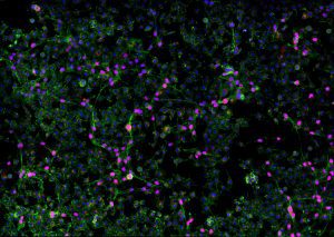
Cellular scattering and variation in the tissue’s oxy and deoxy-hemoglobin concentrations, for example, can be imaged by recording spatial-temporal variations in light absorption as well as the scattering properties of tissue. Diffuse optical tomography is important for preclinical research, neurology, oncology, and surgery. It is sometimes known as near-infrared optical tomography or fluorescence diffuse optical tomography.
How Diffuse Optical Tomography Works
Diffuse Optical Tomography works by differentiating between the optical properties of different biological substances and using them to map differences within the target tissue. Intracellular and extracellular fluids may have different refractive indices, for instance, as do subcellular components (think mitochondria and nuclei). This leads to differences in scattering coefficient and g-factors in different parts of tissue.
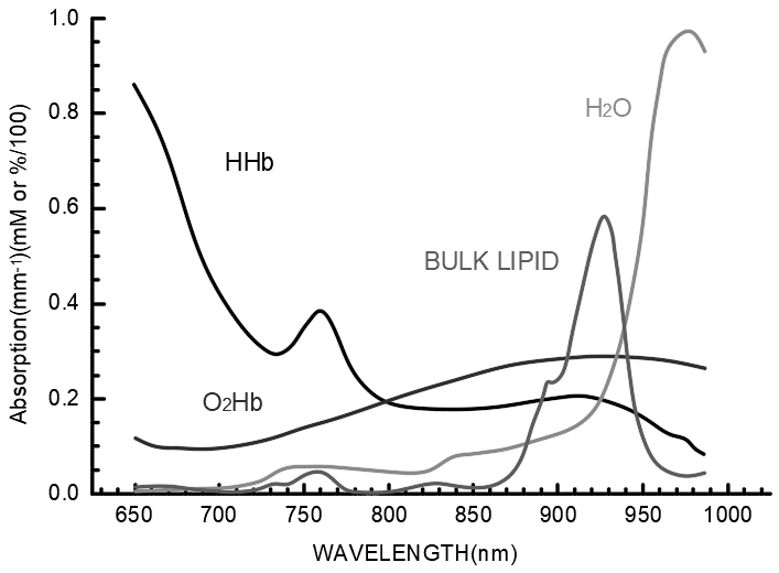
This image shows the varying absorption and scattering coefficients of some important chromophores
Differences in the concentration of various chromophores will also lead to different absorption and scattering coefficients. By measuring transmitted and reflected light on the surface of the tissue, a profile of the inside of the tissue can be determined using reconstruction algorithms. Time correlated single photon counting can also be used to quantify fluorescence lifetimes. These methods enable researchers and medical practitioners to get an inside view of the oxygenation of brain tissue, for instance, or of the specific composition of cancer and healthy cells in breast tissue.
The equipment needed for diffuse optical tomography depends on which methodology is used. The three primary types include time resolved (time domain) systems, frequency-domain devices, and steady-state domain instrumentation. Time domain systems are based on short light pulses, frequency domain devices bathe the tissue in a continuous wave of sinusoidally modulated light, and steady-state domain instrumentation utilizes a constant, steady stream of light.
Fluorescence diffuse optical tomography (FDOT) relies on the use of fluorescent contrast agents, but other applications of DOT may rely only on naturally occurring chromophores.
Advantages and Limitations
Diffuse Optical Tomography (DOT) is particularly useful in clinical and research applications because it is non-ionizing and non-invasive. Equipment needed can be made portable, enabling bedside use, and it is simpler and lower cost than many other imaging techniques. It is also a high-speed technique that offers immediate results.
That said, diffuse optical tomography does have its limitations. Chief among them is the low spatial resolution as compared with other imaging techniques. A DOT image will never be as high resolution as that achieved through magnetic resonance imaging (MRI), but the portability and simplicity of the procedure make it nevertheless extremely useful in clinical and research settings.
Applications
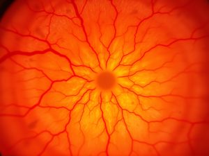
Diffuse optical tomography has been used extensively in studies of the human brain. It can be used in both adult patients and neonates, and can be used to provide on-the-go information about brain activity and oxygenation.
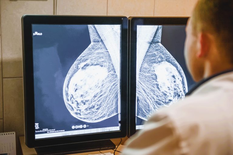
Diffuse optical tomography can also be used for cancer screening, particularly for breast cancer detection. Breast cancer imaging involves creating a map of absorbers like oxy-hemoglobin (HbO2), deoxy-hemoglobin (Hb), water, and lipids. This is instrumental to diagnosis since total hemoglobin concentration is strongly correlated with tumor position.
Another application of DOT is in the imaging of muscles, joints, and peripheral circulation, enabling early flagging of problematic oxygenation or circulation.
Fiber optic arrays on breast tissue enable diffuse optical tomography to detect tumors
Optics for Diffuse Optical Tomography at Avantier
At Avantier we produce a wide variety of custom optics for medical and research applications. Our teams have extensive experience with the unique requirements and challenges of medical optics, and we are available to work with you to design and produce the custom optics that can take your practice or research to the next level. Contact us today to place your initial order or schedule a consult.