
Finite conjugate tube lenses are optimized for fixed-distance imaging, offering precise magnification and strong aberration control. They are ideal for microscopy and inspection but require careful alignment.
Finite conjugate tube lenses are optimized for fixed-distance imaging, offering precise magnification and strong aberration control. They are ideal for microscopy and inspection but require careful alignment.
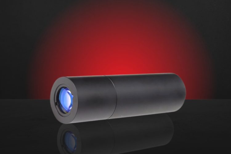
Learn how Avantier partnered with a global leader in medical laser technology to replace outdated systems with advanced germanium lenses for critical applications like laser surgery and thermal diagnostics.

Key Takeaways ISO 10110 is the global standard for representing optical components in technical drawings, ensuring clear communication between designers and manufacturers. It defines specifications for surface form, material quality, surface texture, coatings, tolerances, and alignment—critical for precision lens design. By standardizing symbols and notations, it minimizes errors, improves quality, and supports international collaboration. Adhering […]

Key Takeaways: Thermal management in space optics Thermal management in space optics ensures stable performance across the wide thermal variations typical of orbital environments—using passive techniques like matched-CTE materials, athermal optical design, and radiative shielding. These strategies prevent focus shift, wavefront distortion, and mechanical misalignment without adding mass or complexity. From CubeSats to deep space […]
Key Takeaways Projects in aerospace, biomedical imaging, and other high-precision sectors often hinge on expert optical engineering. Warning signs you need this expertise include: when optical performance is mission-critical, prototypes rely on trial-and-error, off-the-shelf optics fail specs, integration issues degrade performance, or timelines can’t support in-house hires. Skilled optical engineers apply advanced modeling, custom design, […]
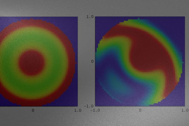
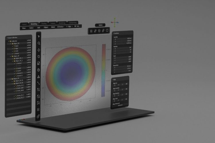
Introduction to SSI Subaperture Stitching Interferometry In the evolving landscape of optical systems manufacturing, test results with high measurement accuracy are critical—especially for large aperture and aspherical surface components used in technologies such as astronomical telescopes and EUV lithography. Conventional interferometers struggle to capture precise full aperture measurements due to limitations in range and aberration […]
Key Takeaways Meeting strict wavefront error (WFE) budgets is critical for space telescope optics, ensuring nanometer-level imaging accuracy and mission reliability. This article details practical strategies for managing WFE contributions from surface figure, alignment, coatings, and thermal effects. Advanced metrology techniques, such as phase-shifting interferometry and stitching profilometry, support precise wavefront measurement. By integrating custom […]
Custom high-NA objective lenses achieve ultra-precise UV excitation and imaging in ion trap experiments, boosting fluorescence capture and quantum accuracy.

Key Takeaways Optical sensors for ADCS—including star trackers, sun sensors, and Earth horizon sensors—provide the high-precision attitude knowledge required for CubeSats and small satellite platforms. Avantier delivers space-qualified optical components engineered for SWaP-constrained missions, offering compact, radiation-hardened systems that maintain performance under vibration, thermal cycling, and radiation exposure. Miniaturized, athermally stabilized optics and lightweight optical […]

Laser-Induced Damage Threshold (LIDT) defines the laser energy level that causes permanent optical damage. This guide explains mechanisms, testing methods, and key factors for safe, optimized laser system design.