Key Takeaways
- Fourier Ptychographic Microscopy (FPM) is an innovative computational imaging technique that overcomes traditional limitations in microscopy by offering both high resolution and a wide field of view.
- Key advances, including ESA-FPM, REFPM, and LRA-piFP, enhance speed, resolution, and robustness.
- Applications span digital pathology, drug screening, 3D imaging, and metrology.
- Manufacturing challenges include LED precision, optical aberration, and sample thickness limitations.
Super-resolution Fourier Ptychographic Microscopy (FPM)
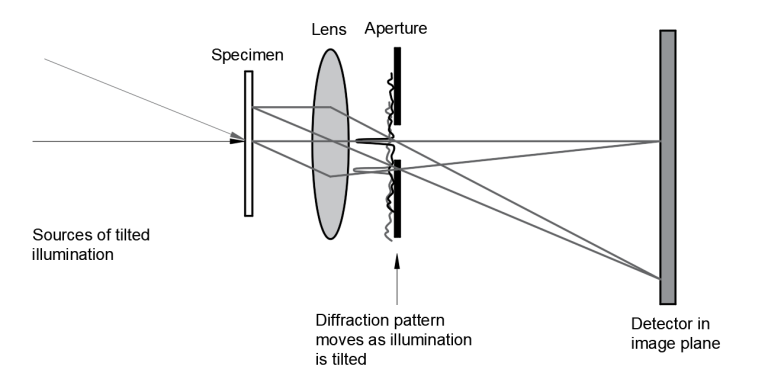
FPM addresses the fundamental trade-off between resolution and field of view in conventional microscopy by combining principles of structured illumination, ptychography, and phase retrieval. This technique enables high resolution, wide field imaging using low numerical aperture, low magnification objective lenses.
Super-resolution Fourier Ptychographic Microscopy Principles
FPM utilizes an array of programmable LEDs for illumination from different angles, expanding the frequency domain bandwidth by overlapping pupil functions. Key components include:
- LED array illumination: Provides angularly varying illumination to capture multiple low-resolution images of the sample from different incident angles.
- Low-NA objective lens: Captures wide field-of-view images at low resolution, which are later computationally enhanced.
- Digital camera: Records the series of low-resolution intensity images corresponding to different illumination angles.
- Computational reconstruction algorithms: Combines the captured low-resolution images in the Fourier domain to synthesize a high resolution image with both amplitude and phase information.
The reconstruction process alternates between spatial and Fourier domains, applying constraints to produce a high-resolution, wide-field complex sample image.

Advantages of Super-resolution Fourier Ptychographic Microscopy
- High Resolution and Wide Field of View (FOV): This technology combines the advantages of both high-magnification and low-magnification imaging, allowing for detailed views across large sample areas.
- Resolution Enhancement: It utilizes computational techniques to improve resolving power beyond the physical limits of the optical system.
- Phase Retrieval Capabilities: This feature enables the extraction of phase information from samples, allowing for the visualization of transparent structures without the need for staining.
- Digital Aberration Correction: The system can computationally correct optical imperfections, enhancing image quality without requiring modifications to physical lenses.
Recent Advancements
- Efficient Synthetic Aperture FPM (ESA-FPM) reduces the number of required raw images, significantly decreasing acquisition time.
- Resolution-Enhanced FPM (REFPM) pushes the resolution limits even further by incorporating advanced optical designs.
- Low-Rank Approximation FPM (LRA-piFP) improves reconstruction robustness in the presence of noise and environmental perturbations.
Applications
- Digital pathology: Enables rapid, high-resolution scanning of large tissue samples for diagnostic purposes.
- Drug screening: Facilitates high-throughput analysis of cellular responses to pharmaceutical compounds.
- Three-dimensional imaging: Allows for the reconstruction of 3D structures from 2D image data.
- Label-free imaging: Provides contrast in transparent samples without the need for staining or fluorescent markers.
- Metrology and scientific research: Offers high-precision measurements for various scientific and industrial applications.

Manufacturing Challenges
- LED array precision: Requires extremely accurate positioning and control of multiple light sources.
- High precision motorized stage requirements: Necessitates nanometer-level positioning accuracy for sample or optics movement.
- Illumination brightness consistency: Demands uniform light output across all LEDs in the array.
- Optical aberration minimization: Requires high-quality optics to reduce inherent system aberrations.
- Sample thickness limitations: Imposes restrictions on the thickness of samples that can be effectively imaged.
Case Study: Super-Resolution Imaging of Biological Cells
Experimental Setup
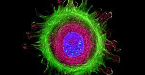
– Sample: Fixed HeLa cells stained with fluorescent dyes
– Microscope: Standard brightfield microscope modified with LED array
– Illumination: Programmed LED array for structured illumination
– Reconstruction: Fourier ptychographic phase retrieval algorithms
Findings
- Resolution Enhancement: Achieved ~0.6 µm spatial resolution
- Phase Contrast Imaging: Revealed detailed phase information
- Large Field of View: 10x larger than confocal systems
- Cost and Time Efficiency: Significantly more affordable and faster
- Applications: Detailed visualization of sub-cellular structures
Challenges
- Computational Load: Required GPU-based processing
- Alignment Sensitivity: Precise calibration needed
- Noise Handling: Preprocessing steps required
Impact and Future Directions
– Potential for transformative applications in cancer research, drug development, and pathogen detection
– Future focus on live imaging, machine learning integration, and hardware optimization
Leading Institutions in FPM Research
- California Institute of Technology (Caltech)
- Chinese Academy of Sciences (CAS)
- University of Connecticut
- Howard Hughes Medical Institute
- National Science Foundation (NSF)
Fourier Ptychographic Microscopy Advancing Imaging Innovation
Fourier Ptychographic Microscopy represents a significant advancement in microscopy techniques, offering high resolution, wide field imaging with phase information. Despite manufacturing challenges, its potential applications in various fields, particularly in digital pathology, make it a promising technology for future research and development.
- https://www.nature.com/articles/s41598-017-09090-8
- https://medicalxpress.com/news/2024-06-feature-domain-fourier-ptychographic-microscopy.html
- https://pmc.ncbi.nlm.nih.gov/articles/PMC10887115/
- https://pmc.ncbi.nlm.nih.gov/articles/PMC4369155/
- https://clpmag.com/diagnostic-technologies/anatomic-pathology/microscopy/fourier-ptychographic-microscopy-may-transform-digital-pathology/
- https://pubmed.ncbi.nlm.nih.gov/38391937/
- https://phys.org/news/2022-10-rapid-full-color-fourier-ptychographic-microscopy.html
GREAT ARTICLE!
Share this article to gain insights from your connections!


