Biomedical applications benefit from advanced filters in Raman spectroscopy to analyze scattered light during neurosurgery.
Laser blocking filters like notch or edge pass filters enhance signal clarity by isolating inelastic scattered light from Rayleigh scattering.
Raman spectroscopy aids in real-time tissue analysis, distinguishing between healthy tissue and pathological conditions.
Avantier offers custom filters optimized for biomedical research, improving diagnostic capabilities in fields like osteoporosis.
Raman Spectroscopy Advancements in Biomedical Applications
Advanced Raman filters can be used in Raman spectroscopy to provide real-time data on biological tissue. They have enormous potential in medicine, research, and biomedical engineering, and can be adapted to a wide variety of different applications.
A basic Raman spectroscopy setup will give you just that: basic information. But what if you are working in biomedical and bio analytical applications and need something more? Advances in Raman spectroscopy involve using a precise configuration and carefully chosen filters to maximize the amount of information this technique can provide. The appropriate laser wavelength and ideal filter will be different for different types of biological samples, so it’s important to do your research and determine what parameters are right for your application.
We’ll go into this in more detail in a moment. But before we begin, let’s quickly step back. How does Raman spectroscopy work, anyway?
Before We Begin: How does Raman Spectroscopy Work?
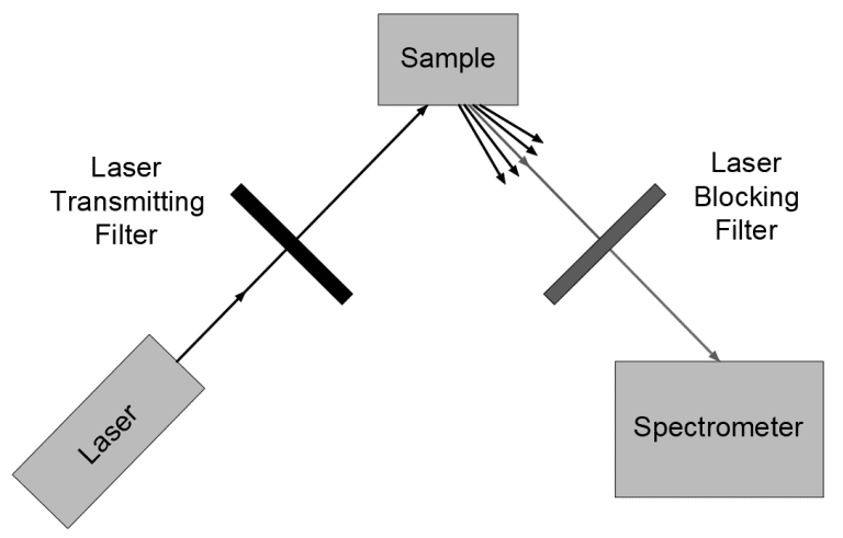
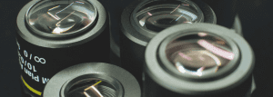
Typically Raman spectroscopy is run by illuminating a sample with a laser beam and observing the scattered radiation that results. This scattered light can be categorized into two sets: Rayleigh scattering, at the laser wavelength, and inelastic scattered light. The inelastic scattered light is what tells us something about the sample, but the Rayleigh scattering has much higher intensity. A laser blocking filter (either a notch filter, edge pass filter, or band pass filter) is used to separate the Rayleigh scattering from inelastic scattered light, the ramen signal.
Just what is this scattered light, and how can it make a difference to human health? We call it Raman scattering, and, as it turns out, a good signal can tell us what molecules are present in a sample and even what the intermolecular bonds look like.
In the past, holographic gratings and multiple dispersion stages were used to separate out the Raman scattering, which then might be collected by photomultipliers. Today, notch or edge filters are the optics of choice to eliminate unwanted Rayleigh scatter and provide a clear signal.
Let’s look at two real-world examples of advanced spectroscopy at work in the biomedical sciences, one in real-time neurosurgery, and another in giving medical practitioners and researchers a better understanding of the process and progression of osteoporosis.
Raman Spectroscopy for Real Time Tissue Analysis in Neurosurgery
It’s real-time and it’s non-destructive. It can be used to view tissue at the surface, but also provide a chemical fingerprint of cells many layers deep. All that makes Raman spectroscopy ideal for real time tissue analysis during neurosurgery, as part of the ‘visual eye’ that guides the knife.
Residual tumor—- a small amount of cancerous cells left after the bulk of a tumor has been cut away— is one of the main causes of neurosurgery failures. That’s why it is so important to be able to differentiate between healthy and cancerous tissue while the surgery is ongoing. Raman spectroscopy has the potential to do this, differentiating between healthy tissue, pathological tumors and necrosis. Raman techniques like CRS, CARS, or SRS microscopy can be used to provide a stronger signal and quicker imaging times.
Raman Spectroscopy for Osteoporosis Diagnosis
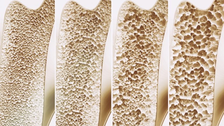
Osteoporosis diagnosis is another area in which Raman spectroscopy shows promise. Osteoporosis results in reduced bone quality, bone fragility, and low BMD, but historically there has been no medical diagnostic method that can be used to assess the bone quality of a living subject.
Raman spectroscopy has the potential to step into that gap. Testing on mice has shown that Raman spectroscopy can provide a real time view of the changes in carbonate/phosphate ratio and mineral/matrix ratios that occur when osteoporosis sets in. It may not be long till these techniques enable a better understanding of how osteoporosis affects human health and how biomedical sciences can reverse or stop the bone deterioration involved.
Raman spectroscopy can be used to gauge bone density and bone quality, critical markers of osteoporosis
Selecting Optimal Optical Filters for Your Application
At Avantier, we produce custom Raman edge filters, Bandpass filters and notch filters optimized for blocking the laser line and transmitting anti stokes Raman scattering. We can also supply laser line filters; these filters transmit the exact wavelength of laser light needed to illuminate the sample while blocking other radiation that might potentially degrade the signal-to-noise ratio.
Our engineers and designers are available for consultation and can help you determine the ideal wavelength ranges, optical density, and type of filter for your application. We can also work with you to determine the ideal configuration and angle of incidence for your system. Contact us today for your next project.