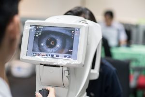
Optical medical imaging is transforming diagnostics, surgery, and research.
Diffusive optical imaging (DOI) uses NIR spectroscopy to study soft tissue, while ballistic optical imaging detects unscattered photons for high-resolution imaging, offering better optical quality than MRI or ultrasound.
Photoacoustic imaging combines optical and ultrasonic methods for deep, detailed scans. Positron emission tomography (PET) tracks radioactive tracers for real-time molecular insights, though radiation precautions are needed.
Avantier offers custom optical solutions for advanced medical applications.
Optical Medical Imaging
Whether your focus is medical diagnostics, surgery, clinical care, or biomedical research, optical medical imaging can play a transformational role in your practice. Recent advances in optical research are pushing the bar on medical imagery, and today’s practitioners have tools at their fingertips that could only have been dreamed of a few decades ago.
Here we’ll look at two basic types of optical medical imagery that are both seeing transformative advances: diffusive optical imaging and ballistic optical imaging. We’ll also look briefly at nuclear imaging and a special hybrid type of medical imaging: photoacoustic imaging.
Diffusive Optical Imaging
Diffusive optical imaging (DOI) involves using NIR spectroscopy or fluorescence-based imaging to elucidate the optical properties of biological tissue. Most useful for soft tissue, a strongly diffusive media, this type of imaging can be used to determine the real-time concentration of key chromophores in the tissue through measurement of absorption and scattering coefficients. For instance, DOI can be used to monitor changes in oxygenated or deoxygenated hemoglobin, or the redox states of cytochromes.
One possible setup for DOI involves time-resolved excitation and detection schemes. A multiple picosecond diode laser sends pulses to the tissue to be imaged. As the laser pulses exit the tissue, temporal profiles are recorded using time-correlated single photon counting (TSPC). The shape of the pulses, analyzed with physical models of the absorption and scattering properties of the tissue, is used to generate images.
When diffusive optical imaging is used to create 3D images it is called diffusive optical tomography (DOT). Breast cancer imaging, stroke detection, muscle functional studies, and brain functional imaging are just a few of the fields in which DOI and DOT are applied on a regular basis.
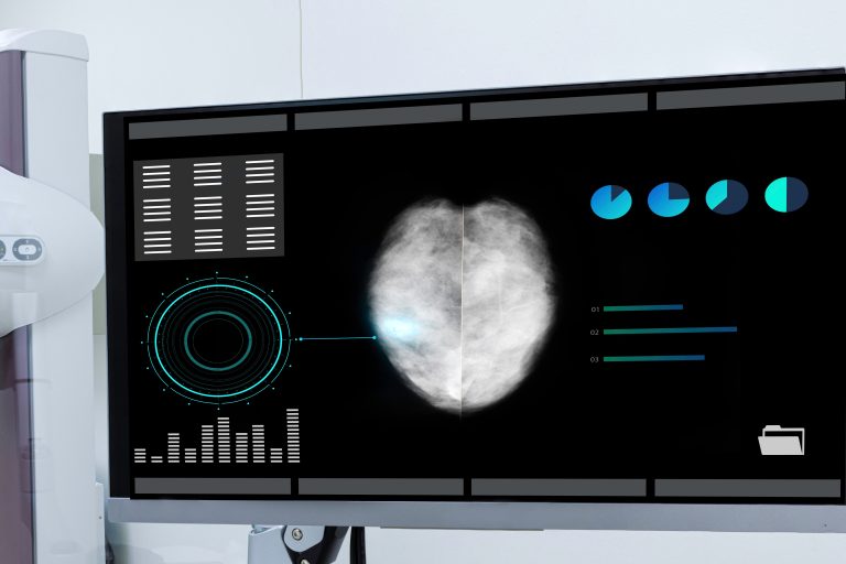
A mammography captured by diffusive optical imaging, a noninvasive form of optical medical imaging.
Ballistic Optical Imaging
Ballistic optical imaging produces high-resolution images of biochemical tissue using techniques like optical coherence tomography (OCT) and ballistic scanners featuring ultrafast time gates. Unlike diffusive optical imaging, this type of imaging relies on detecting photons that travel through the tissue in a straight line without being scattered or ‘diffused’.
Although ballistic optical imaging technology has a limited working depth, the images it produces can have better optical qualities than images produced by alternate imaging technology like magnetic resonance imaging (MRI), ultrasound, or X-ray. When the procedure is used for deeper tissue, image processing techniques are used to reconstruct higher-quality images from multiple raw ballistic images.
Photoacoustic Medical Imaging
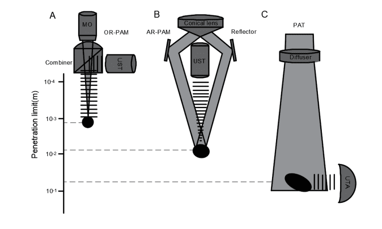
Photoacoustic imaging (also known as optoacoustic imaging) is a revolutionary new type of medical imaging that combines high contrast optical imaging with deep penetrating ultrasonic imaging. The result is high resolution images that provide both structural and functional information.
Non-invasive and non-ionizing, the method produces diverse endogenous and exogenous contrast and good imaging depth. Types of photoacoustic medial imagery include photoacoustic tomography (PAT) and photoacoustic microscopy (PAM).
Different configurations for photoacoustic medical imagery enable different penetrations and resolutions.
Positron Emission Tomography
Positron emission tomography (PET) works in a way fundamentally different from the other imaging modalities we’ve looked at here. A type of nuclear imaging, PET is based on tracking a radio tracer, an injectable radioactive chemical that is absorbed at different rates in different tissues. High absorption of the radio tracer signals diseased or cancerous cells, allowing medical professionals to diagnose issues that might be hard to identify with other imaging methods.
Unlike computer tomography (CT) and magnetic resonance imaging (MRI), a PET scan is a imaging procedure that focuses on real-time molecular activity and processes. It may be performed at the same time as other imaging techniques for high resolution 3D images. While the method forms an important part of medical diagnostics, the procedures are time consuming, and precautions around the use of radioactive materials used have limited the ease of use of this method.
Optical Medical Imaging at Avantier
At Avantier, we produce high performance custom optics for cutting-edge medical research, biomedical engineers, and clinical applications. Whether you know exactly what optic you require or need assistance to design an imaging system for your medical application, we can work with you.
Our experienced team of optical designers and engineers is available to put their expertise to work for your application, and we are proud of our track record of providing custom optical systems to satisfied clients worldwide. Contact us today to set up an initial consult or inquire about the custom order process.