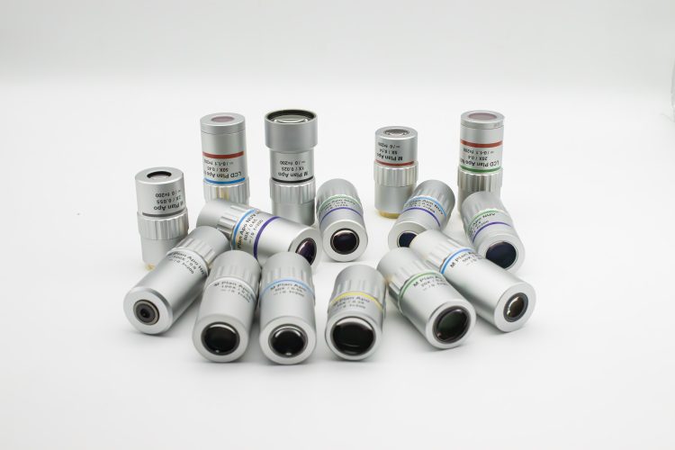
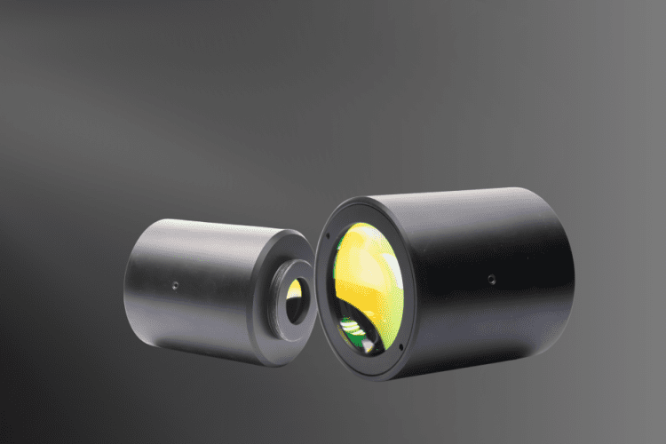
SWIR Camera Surveillance

Key Takeaways SWIR Camera Surveillance uses SWIR light to achieve superior SWIR imaging in conditions where visible light fails. SWIR light penetrates fog and smoke better than visible light, providing clearer images. SWIR imaging is unaffected by heat haze and atmospheric variations, ensuring consistent quality. SWIR cameras use sensors like InGaAs or MCT, often integrating with existing systems with minimal changes. SWIR Camera Surveillance: Eliminating Haze There’s a deep fog over everything, and you can’t see your hand when you hold it in front of you. The perfect night for a thief to sneak into your factories, make out with whatever they can carry, and get out the same way they came— or is it? Not if you have SWIR camera surveillance. While visible light cannot penetrate well in water vapor, SWIR light has no problem that way. A SWIR camera can produce clear, high resolution images even when the human eye is unable to make heads or tails of anything. Here we’ll look at just what SWIR can do and how it does it.  SWIR camera surveillance makes it possible to get a clear view of a foggy landscape. The Foundations of SWIR Camera Surveillance SWIR is short for short wave infrared and is the part of the spectrum with wavelength between 0.9 and 1.7 microns. Photons of SWIR light are absorbed and reflected from an object in a way analogous to visible light. It enables a high dynamic range and good contrast in imaging. But this light is invisible to the human eye, and the silicon sensors used for visible light imaging don’t work with SWIR. Dedicated SWIR sensors (typically made of InGaAs or MCT) are used instead.  SWIR lighting can illuminate an industrial complex at night while maintaining the appearance of darkness. Outdoors, natural SWIR (‘night glow’) is present under nearly all weather conditions, both day and night.   Night glow is a type of atmospheric radiance that allows SWIR camera surveillance in even the darkest nights. Why SWIR Camera Surveillance? We’ve touched briefly on why short wave infrared SWIR camera surveillance is so important to a wide range of applications, but let’s look at it in more detail now. The importance of SWIR imaging lies in its wavelength. Since SWIR light has a shorter wavelength than visible light, it isn’t scattered by the microparticles of fog and smoke. This makes SWIR imaging more effective in such conditions compared to visible light. Heat haze and atmospheric temperature variances can cause havoc with thermal imaging. However, these factors do not affect SWIR imaging. This makes SWIR imaging ideal for ensuring clear images at any time, whether it is night or morning. It also works effectively regardless of the atmospheric conditions.  Visible LWIR SWIR SWIR camera surveillance can provide clear images even when both visible and LWIR imaging fails. Is that the only reason major players in industry, defense, and security are turning to SWIR camera surveillance? No, there are more.  One benefit of SWIR vision systems is that no visible illumination is needed. As mentioned in the last section, a powerful beam of SWIR light can illuminate a setting that looks dark to everyone using more traditional imaging equipment. A SWIR sensor can also be used to locate lasers and beacons, making it helpful in keeping track of mobile teams or for military IR laser spotting and tracking.  In biometrics, SWIR can be used to distinguish between fake hair and human hair. This enables easy identification of individuals who might be attempting to disguise their identity. Human hair will appear light, while fake hair shows up as dark. There’s nothing easier than picking out a person who is carrying around a head of hair that was manufactured in a factory.  SWIR Imaging Equipment The InGaAs sensors used for SWIR imaging are manufactured from indium gallium arsenide. These sensors can detect light radiation ranging from 550nm to as high as 2.5μm. An alternative to InGaAs is MCT, or mercury-cadmium detector, which can detect into the long wave infrared LWIR region. The benefit of MCT is that it is ‘tunable’. The optical absorption wavelength changes based on the cadmium concentration. This allows the amount of cadmium (Cd) to be chosen to create a sensor that absorbs optimally in a given region. There is a big downside to MCT sensors, however— they must be cryogenically cooled.  SWIR radiation travels through glass in much the same way as visible light. This means that standard camera lenses, mirrors, and other optics can be used in short wave cameras.  If you’d like to switch your existing surveillance system over to SWIR you may need to make only small modifications.  It may take less than you think to set up your new SWIR camera surveillance system. Custom SWIR Camera Surveillance At Avantier, we’re all about producing custom optics that enable our customers to reach their performance goals, no matter what the application. Whether you need a SWIR camera with high frame rates and low exposure time, a SWIR vision system for an unmanned vehicle, or SWIR imaging equipment for a research project, we can assist you. We provide the optical components and lens assemblies necessary for your specific needs. Just let us know your requirements, and we’ll tailor our solutions accordingly. Related Content

Read more
The Complete Guide to Microscope Objective Lens
the complete guide to microscope objective lens header

Product Highlights: A microscope magnifies objects, revealing details not visible to the naked eye. It can project the image to the eye or a camera for observation or recording. Avantier’s diverse lineup, featuring achromatic, semi-apochromatic, and apochromatic options, ensures superior imaging quality. With magnifications ranging from 1X to 100X and numerical apertures up to 1.45, Avantier caters to a wide range of microscopy needs.  Avantier excels in specialized objectives like polarizing, phase contrast, and metallurgical objectives. Custom solutions by Avantier extend capabilities into UV, NUV, and NIR regions, to push the boundaries of precision microscopy for research, industry, and medical diagnostics. The Complete Guide to Microscope Objective Lens​ Table of Contents What is a Microscope Objective Lens? Microscope objective lenses, vital optical elements in microscopy, enable precise observation of specimens. Objective lens manufacturers offer a wide range of objective designs for specific needs: high power for detailed observation, scanning for broader views, oil immersion for high-resolution imaging, and long working distance for manipulation without compromising quality. Those objectives are designed with advanced construction techniques for high performance objectives with a spring loaded retractable nose cone assembly that protects the front lens elements and the specimen from collision damage. Adding to these features, long working distance objectives allow ample space between the lens and the specimen, facilitating the manipulation of samples without compromising image quality. Infinity correction objectives utilize infinity-corrected optical systems, providing flexibility and compatibility with various microscopy accessories. Numerical aperture, magnification, optical tube length, degree of aberration correction, and other important characteristics are typically imprinted or engraved on the external portion of the barrel for easy reference. These specifications help researchers select the appropriate objective for their experiments, ensuring optimal performance and total magnification when combined with the ocular lens. Specifications like numerical aperture and magnification are typically labeled on the barrel for easy reference. These lenses are indispensable in scientific research providing high powered optics essential for research. In the following content, we delve intensively into the various components and features of microscope objective lenses, exploring their construction, functionality, and specialized designs that enable researchers to gain deeper insights into the microscopic world. Components of a microscope A microscope is an optical device designed to magnify the image of an object, enabling details indiscernible to the human eye to be differentiated. A microscope may project the image onto the human eye or onto a camera or video device.  Microscopes are usually complex assemblies that include an array of lenses, filters, polarizers, and beamsplitters. Illumination is arranged to provide enough light for a clear image, and sensors are used to ‘see’ the object. Although today’s microscopes are usually far more powerful than the microscopes used historically, they are used for much the same purpose: viewing objects that would otherwise be indiscernible to the human eye.  Here we’ll start with a basic compound microscope and go on to explore the components and function of larger more complex microscopes. We’ll also take an in-depth look at one of the key parts of a microscope, the objective lens. In many microscopes, backlight illumination is favored over traditional direct light illumination due to the latter’s tendency to over-saturate the object under inspection. One specific backlight illumination technique employed in microscopy is Koehler illumination. This method involves flooding the object with light from behind using incident light from a source like a light bulb (see Figure 2). Koehler illumination utilizes two convex lenses, the collector lens and the condenser lens(or called field lens) , to ensure even and bright illumination on both the object and image planes. This design prevents imaging the light bulb filament, a common issue with direct light illumination. Backlight illumination is also commonly referred to as brightfield illumination. Figure 1 For brightfield illumination to be effective, there needs to be a variation in opacity across the object. Without this variation, the illumination creates a dark blur around the object, resulting in an image with relative contrast between the object’s parts and the light source. Typically, brightfield illumination allows clear visualization of each part of the object unless it is extremely transparent. In cases where transparency hinders feature distinction, darkfield illumination becomes useful. Darkfield illumination directs light rays obliquely onto the object, avoiding direct entry into the objective. Despite this oblique angle, the rays still illuminate the object plane. The resulting darkfield illumination image achieves high contrast between the transparent object and the light source. In a darkfield setup, a light source forms an inverted cone of light that blocks central rays but allows oblique rays to illuminate the object (see Figure 3). This design effectively forces light to illuminate the object without entering the optical system, making darkfield illumination particularly suitable for transparent objects. In contrast, no rays are blocked in a brightfield illumination setup. Figure 2 Figure 3 Epi-illumination, a third form of illumination employed in microscopy, generates light from above the objective. This setup replaces the need for a Koehler illumination configuration, as both the objective and the epi-illumination source contribute to the illumination process. The compact structure  of epi-illumination is a significant advantage, as the objective serves as a primary source for a considerable portion of the illumination. Figure 4 provides a depiction of a frequently used epi-illumination setup, particularly common in fluorescence applications. Figure 4 Compound Microscope While a magnifying glass consists of just one lens element and can magnify any element placed within its focal length, a compound lens, by definition, contains multiple lens elements. A relay lens system is used to convey the image of the object to the eye or, in some cases, to camera and video sensors. A basic compound microscope could consist of just two elements acting in relay, the objective and the eyepiece. The objective relays a real image to the eyepiece, while magnifying that image anywhere from 4-100x.  The eyepiece magnifies the real image received typically by another 10x, and conveys a virtual image to the sensor.  There are two major specifications for a microscope: the magnification power

Read more