
Optical coatings, including HR and dielectric coatings, enhance the reflectance and transmittance properties of optical components.

Optical coatings, including HR and dielectric coatings, enhance the reflectance and transmittance properties of optical components.
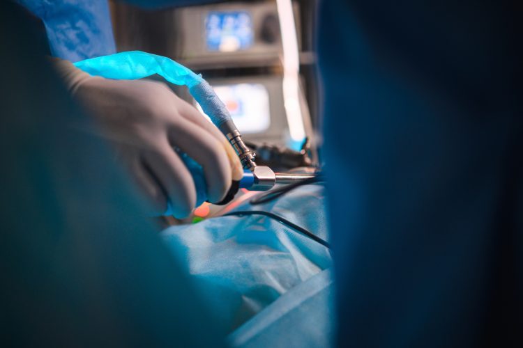
Customized rod lenses in endoscopy facilitate light transmission for precise visualization of internal structures.

The optimization of parabolic mirror telescopes ensures exceptional image quality for astronomical observation.

Infrared imaging requires wavelength-specific Anti-reflective Coatings and Bandpass filters for optimal performance.

Microscope objectives, with their varied objective lenses, determine image resolution through their numerical aperture.

Biophotonics, an interdisciplinary field, explores the interrelationship between biology and optics. It encompasses technologies that utilize light to analyze, detect, and manipulate biological information.

Raman spectroscopy is a powerful analytical technique that provides insights into the molecular composition and structure of materials. Raman spectroscopy relies on the interaction between photons and molecular vibrations.

Avantier stands at the forefront of the Micro-Optics industry, offering original design solutions tailored to meet the unique demands of their clients. Learn about the various applications of micro-optics and how Avantier’s custom solutions can meet the needs of your specific project.

Part 1 discussed the principles and customization options available through Avantier to accelerate research in flow cytometry. In Part 2, we will explore the advantages of customized components such as lenses, filters, mirrors, beamsplitters
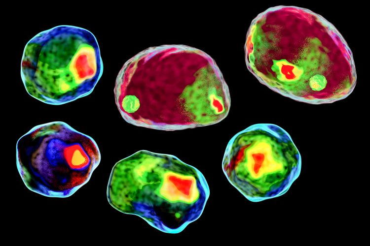
Flow cytometry is a revolutionary technique that enables the comprehensive analysis of cells or particles in a high-throughput manner. By harnessing the principles of hydrodynamic focusing, electronic detection, and optical systems, flow cytometry provides valuable insights into cell populations