Key Takeaways:
- Indirect ophthalmoscopes use a high‑diopter double convex lens as a front lens to create an enlarged inverted real image of the fundus for detailed retinal assessment.
- High‑order aspheric design on one surface of the double convex lens corrects higher‑order aberrations, enhancing image contrast and reducing distortion compared to a purely spherical lens.
- A 40D front lens provides carefully chosen magnification and field of view, supporting efficient eye examinations across a range of patients.
- Precision CNC aspheric machining and tight surface‑figure control (PV < 0.5 µm, eccentricity < 1 arcmin) are critical to maintain imaging performance in clinical use.
- Vis‑NIR broadband antireflection (AR) coatings on the double convex lens minimize reflection losses and support both visible and infrared illumination modes during diagnosis.
Case Study: Double Convex Lens for Indirect Ophthalmoscopes
This case study details the design and manufacturing of a custom double convex lens used as the front lens in an indirect ophthalmoscope, a key instrument for high‑quality fundus examinations. By combining a biconvex geometry with a high‑order aspheric surface, Avantier’s double convex lenses correct higher‑order aberrations, improve image sharpness, and expand the usable field of view for retinal imaging.
For more background on our standard and custom double convex lenses, see our Biconvex / Double Convex Lenses overview.
What Is a Double Convex Lens in This Application?
In geometrical optics, a double convex lens (biconvex lens) has both surfaces bulging outward, giving it positive optical power and the ability to converge incident rays to a real focus. In indirect ophthalmoscopy, the front double convex lens is held in front of the patient’s eye so that light reflected from the fundus forms an enlarged, inverted real image in space, which is then re‑imaged by the ophthalmoscope’s internal lens group onto a sensor or the clinician’s eye.
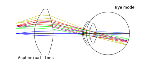
This front lens behaves as a high‑diopter converging element; typical cases of convex lens behavior (object inside, at, or beyond the focal length) map directly to how the fundus image is positioned relative to the device’s imaging plane. In this design, a 40‑diopter high‑order aspheric biconvex lens is used to balance magnification, working distance, and field of view for routine retinal exams.
Learn how this ophthalmic application fits into our broader Medical Optical Components & Lenses portfolio.
Optical Design: From Spherical Lens Limitations to High‑Order Aspherics
Why not a purely spherical convex lens?
In theory, a spherical double convex lens can form the required enlarged fundus image, but spherical aberration and other higher‑order aberrations degrade contrast and limit resolution, especially at high diopter values. This is unacceptable for indirect ophthalmoscopes, where subtle lesions must be visible at the retinal periphery.
To address those limitations, Avantier’s design uses a single‑sided high‑order aspheric surface on the double convex lens. Compared to a traditional low‑order asphere, a high‑order aspheric surface introduces additional degrees of freedom in the sag equation, enabling more precise control of surface curvature and therefore better correction of higher‑order aberrations across the full field.
For a deeper dive into aspheric design principles, see our Complete Guide to Aspheric Lens.
Optical System: Cases of a Convex Lens in Indirect Ophthalmoscopy
In this case of convex lens usage, the double convex front lens is characterized by:
- Diopter: 40D, giving high positive power to create a strongly magnified, inverted real image of the retina at a convenient working distance.
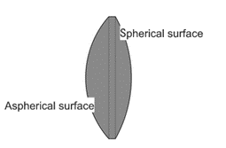
- Geometry: Biconvex (double convex) shape with a single‑sided high‑order aspheric surface, optimized to minimize spherical and higher‑order aberrations.
- Material: H‑K9L (a high‑quality optical glass similar to BK7), chosen for its transmission, homogeneity, and manufacturability for precision ophthalmic lenses.
From a ray‑diagram perspective, this configuration corresponds to the classic positive lens case where the object (fundus) lies beyond the focal length, generating an inverted real image between the lens and the ophthalmoscope’s internal relay lenses. This practical double convex lens diagram closely follows standard convex‑lens imaging theory but incorporates aspheric corrections to enhance real‑world performance.
Specifications of Double Convex Lens
Front Lens Specifications (40D High‑Order Aspheric Double Convex Lens)
Diopter | 40D |
Material | H-K9L |
Shape | Convex lens, single-sided aspherical surface |
Diameter | 40mm |
Thickness | 15mm |
These parameters are chosen to balance magnification, field of view, working distance, and manufacturability in a clinical indirect ophthalmoscope.
Manufacturing Process and Surface‑Figure Control
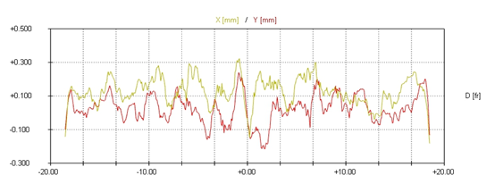
Achieving the required surface accuracy on a double convex lens with a steep aspheric surface is challenging, because small vertex curvature and rapid slope changes are highly sensitive to machining errors. In this case, the aspheric PV (peak‑to‑valley) surface error must be kept below 0.5 µm to avoid noticeable image blur and distortion in retinal images.
Avantier uses a multi‑step CNC aspheric manufacturing workflow:
- The spherical surface is machined first and used as the datum for subsequent clamping and alignment.
- The aspheric surface is then generated using CNC aspheric machining, with a re‑optimized polishing tool specifically designed for the convex aspheric profile.
- Surface accuracy and quality are monitored in‑process (online detection), enabling real‑time correction of form error before final polishing and edging.
- Final centering and edging ensure that lens eccentricity remains below 1 arcmin, minimizing coma and decenter‑induced aberrations in the assembled indirect ophthalmoscope.
This combination of CNC grinding, polishing, and metrology is typical of Avantier’s approach to high‑precision ophthalmic lenses and other medical imaging components.
Coatings and Broadband Imaging Performance
To maximize throughput and compatibility with different illumination modes, the double convex lens is coated with a Vis‑NIR broadband antireflection coating. This coating:
- Reduces surface reflections across the visible and near‑infrared bands.
- Improves contrast and SNR in fundus images under white‑light and NIR illumination.
- Minimizes ghosting and stray light that could mask subtle retinal features.
For related coating capabilities across other optics, explore Avantier’s work on custom optical coatings in our technical and application notes.
Conclusion: Benefits of a High‑Order Aspheric Double Convex Lens
The 40D high‑order aspheric double convex lens described in this case study demonstrates how careful optical design and precision manufacturing can substantially improve indirect ophthalmoscope performance. By moving beyond a simple spherical convex lens and adopting a high‑order aspheric biconvex configuration, the system:
- Corrects higher‑order aberrations and reduces distortion.
- Enhances image sharpness, especially at the retinal periphery.
- Maintains tight control of surface error (PV < 0.5 µm) and eccentricity (< 1 arcmin) for consistent clinical performance.
- Supports Vis–NIR operation, enabling flexible illumination strategies in modern fundus imaging.
Altogether, this double convex lens case illustrates how advanced lens design and manufacturing directly translate into more accurate and efficient ocular assessments, ultimately benefiting both clinicians and patients.
To discuss a custom double convex lens or other ophthalmic lens designs, contact Avantier or review our Aspheric Lens Case Studies for additional examples of high‑precision medical optics.
Related Content: