Optical Coherence Tomography in Medical Imaging: Part 4
Key Takeaways:
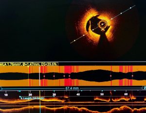
Optical Coherence Tomography (OCT) provides detailed imaging of blood vessels, facilitating diagnosis and treatment of age-related macular degeneration (AMD).
OCTA combines OCT with angiography, offering enhanced visualization of blood vessels and aiding in the diagnosis of retinal diseases.
Key components of OCT systems include a light source, beamsplitter, and detection system, enabling high-resolution imaging of biological tissues.
Different types of OCT systems, such as Time-Domain OCT (TD-OCT) and Spectral-Domain OCT (SD-OCT), offer unique features and imaging capabilities for various medical applications.
Optical Coherence Tomography (OCT) is an advanced imaging technique that has transformed medical diagnostics and research. By providing high-resolution cross-sectional images of biological tissues, OCT enables precise visualization and analysis of various structures, including blood vessels.
With the advent of Optical Coherence Tomography Angiography (OCTA), which combines OCT with angiography, the imaging of blood vessels has become even more detailed and informative. In this article, we will explore the key components and capabilities of OCT systems, their applications, and the impact they have had on diagnosing and managing diseases such as age-related macular degeneration (AMD), macular edema, and more.
At the heart of OCT technology lies the principle of low-coherence interferometry. An OCT system consists of several essential components, including a light source, beamsplitter, reference arm, sample arm, and detection system. The light source emits a beam of light, typically in the near-infrared spectrum, which is split into two beams by the beamsplitter. One beam, known as the optical beam, is directed towards the target tissue or sample, while the other beam serves as the reference beam.
When the optical beam interacts with the tissue, it undergoes scattering and reflection. The back-reflected light from the tissue combines with the reference beam at the beamsplitter, resulting in interference patterns. These interference patterns contain valuable information about the depth or distance of various structures within the sample. By analyzing and processing these interference patterns, cross-sectional images of the tissue can be reconstructed.
One of the key parameters that determine the quality of OCT images is the axial resolution. The axial resolution refers to the ability of the system to distinguish between structures along the depth or axial direction. OCT systems typically achieve high axial resolutions in the range of a few micrometers, enabling detailed visualization of tissue layers and microstructures.
Exploring Different Types of OCT Systems
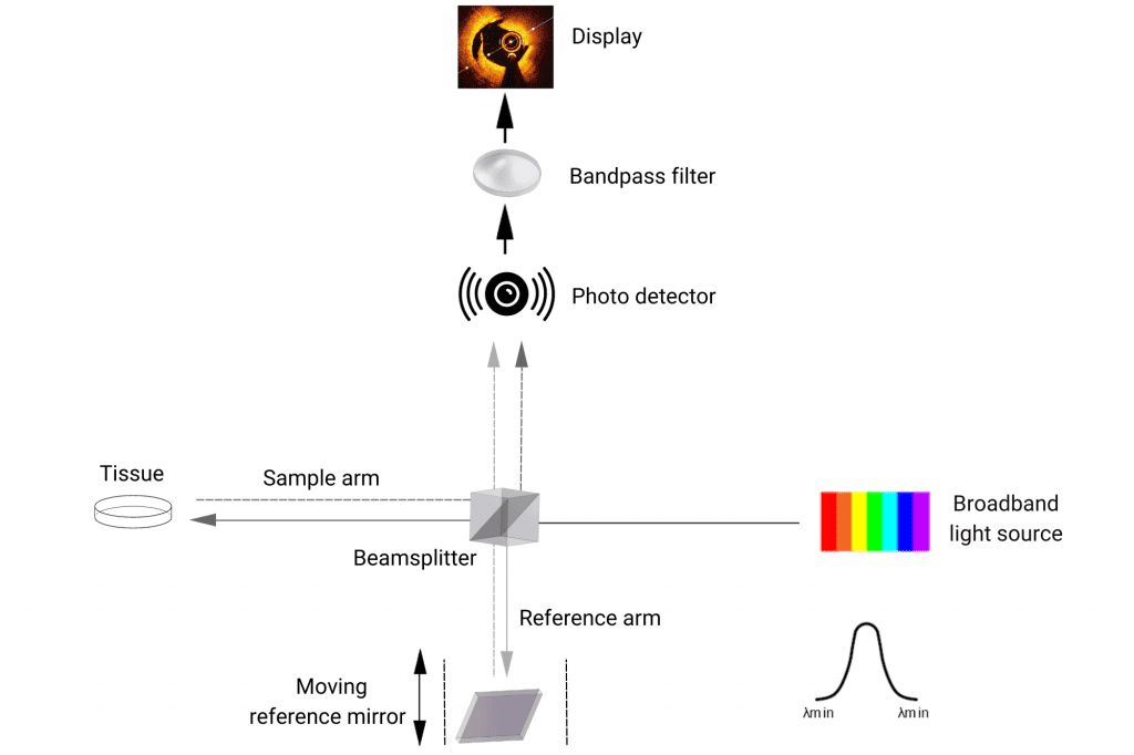
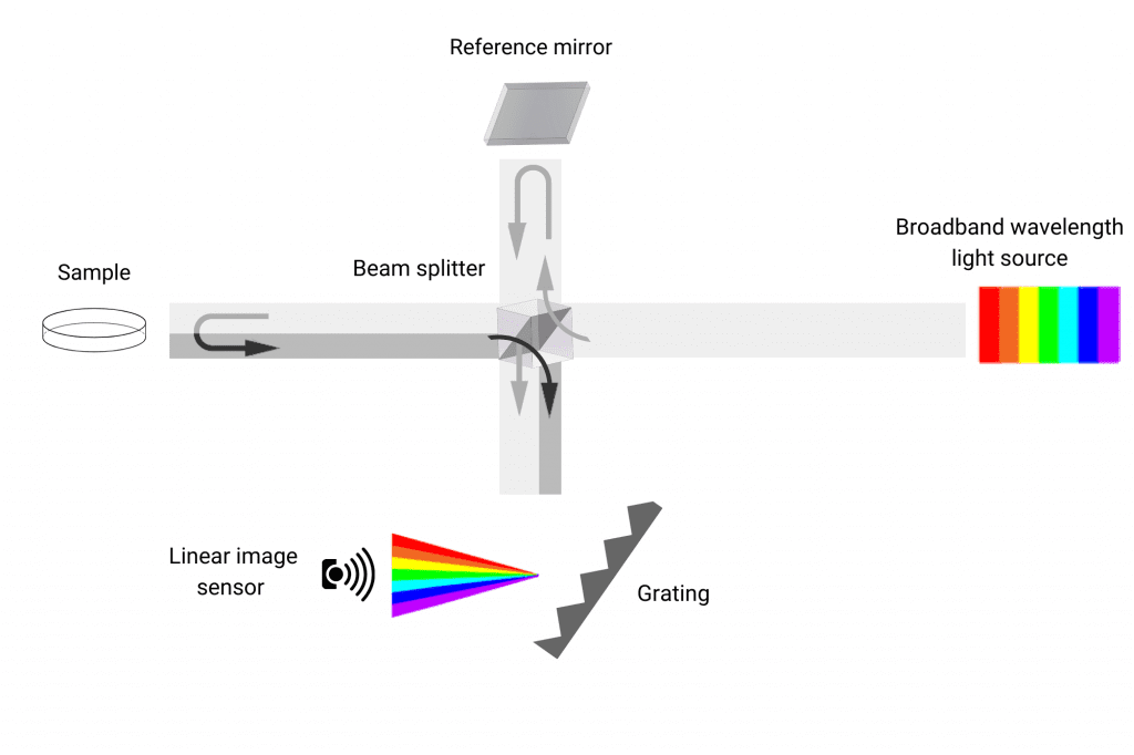
There are different types of OCT systems, including Time-Domain OCT (TD-OCT) and Spectral-Domain OCT (SD-OCT). TD-OCT was one of the earliest implementations and utilized a moving reference mirror to scan the depth of the tissue. SD-OCT, on the other hand, utilizes a spectrometer to simultaneously capture depth information, leading to faster imaging speeds and improved signal-to-noise ratios.
Time-domain OCT system
Spectral-domain OCT system
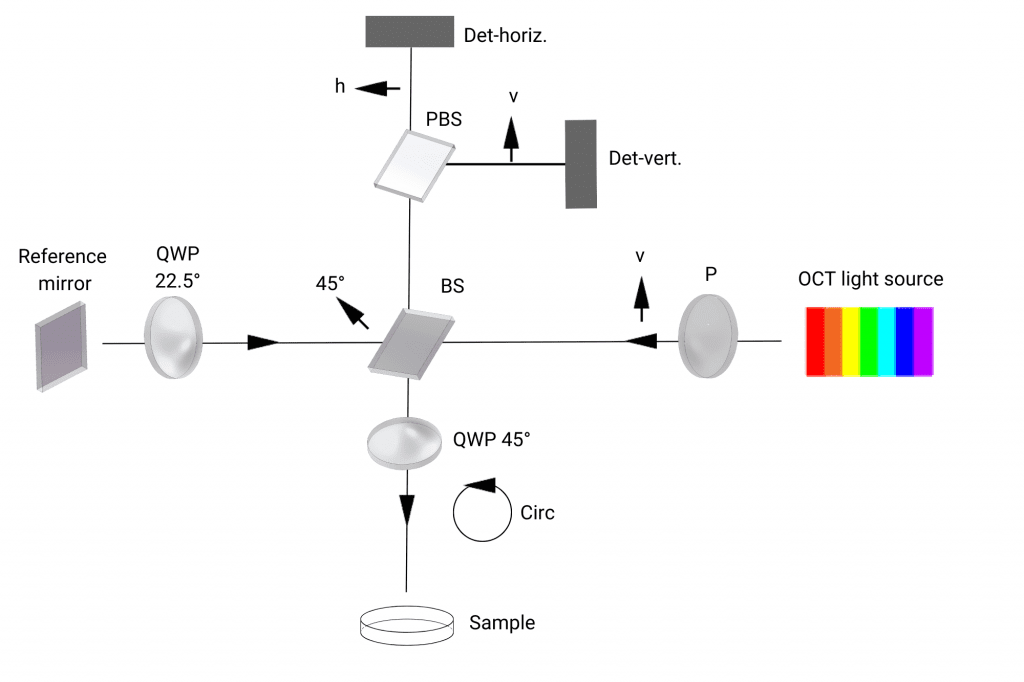
OCT has also evolved to incorporate additional capabilities, such as polarization sensitivity and angiography. Polarization-Sensitive OCT (PS-OCT) utilizes the polarization properties of light to provide information about tissue birefringence and structural characteristics. This allows for the visualization of collagen fibers, nerve bundles, and other polarizing structures within the tissue.
Polarization-Sensitive OCT (PS-OCT)
Unlocking the Potential of OCT Angiography (OCTA) in Medical Imaging and Diagnosis
OCT angiography (OCTA) is a powerful extension of OCT that enables non-invasive imaging of blood vessels. It utilizes the motion contrast of flowing blood cells to create en-face images of the vasculature without the need for contrast agents. By analyzing changes in the OCT signal over time, OCTA can produce detailed maps of the vascular network, allowing for the evaluation of perfusion and identifying vascular abnormalities.
The applications of OCT systems are vast and encompass various medical fields. In ophthalmology, OCT has revolutionized the diagnosis and management of retinal diseases, including age-related macular degeneration (AMD). By providing high-resolution images of the retina and its layers, OCT enables the identification of drusen, retinal pigment epithelial detachments, and fluid accumulation, aiding in the timely diagnosis and monitoring of AMD.