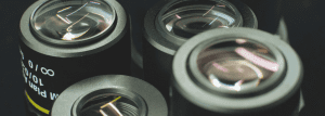
Fluorescent Biological Microscope Objectives: Principles, Functions, and Applications
Key Takeaways
Fluorescence microscope objectives are specialized optical components designed to maximize light collection and resolution ($0.2\mu m$) while minimizing sample damage.
Key design factors include high Numerical Aperture (up to 1.49 for oil), multi-band apochromatic correction for labels like DAPI and FITC, and specialized anti-reflective coatings for weak signal detection.
In the microscopic realm of life science research, fluorescence microscopy has emerged as a pivotal tool for observing cellular structures, tracking molecular dynamics, and deciphering the mechanisms of life. Its primary advantages—specific labeling of biological samples and high-sensitivity imaging—depend heavily on the quality of the optical system.
As the “eyes” of the system, fluorescence biological microscopy objectives directly determine critical performance metrics, including imaging resolution, signal-to-noise ratio (SNR), and light collection efficiency. This article provides a comprehensive analysis of these objectives across three dimensions: design principles, core functionalities, and pivotal applications.
I. Design Principles of Fluorescence Microscope Objectives
The design of fluorescence objectives integrates geometric and physical optics with the unique requirements of biological specimens, such as transparency and sensitivity to light.
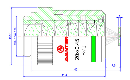
Optical Design of Fluorescent Biological Microscope Lens Designed by Avantier
1. Optimization of Core Optical Parameters
Numerical Aperture (NA): NA is the definitive factor for resolution and light collection, calculated as NA = n ・sin θ. In fluorescence imaging, where signals are often weak, a high NA is essential.
Air objectives: Max NA ≈ 0.95.
Water immersion: Up to 1.2 NA (ideal for live cells).
Oil immersion: Up to 1.49 NA (best for subcellular detail).
Balancing Magnification and Working Distance (WD): High-magnification lenses (e.g., 100x) typically have short WDs (micrometers). Conversely, low-magnification lenses (e.g., 20x) are designed with longer WDs to accommodate thick tissue sections and culture dishes.
2. Targeted Aberration Correction Strategies
Fluorescence imaging is highly sensitive to optical artifacts. Advanced objectives utilize the following:
Chromatic Aberration Correction: Since samples are often multi-labeled (e.g., DAPI, FITC, Texas Red), objectives must achieve Apochromatic (APO) correction to ensure all wavelengths converge on the same focal plane.
Spherical and Coma Correction: By using aspherical lenses and optimized lens groupings, designers minimize image blurring and detail loss caused by light entering at oblique angles.
Field Curvature and Distortion: Symmetrical lens designs ensure the entire field of view (FOV) is flat and uniform, which is vital for large-scale tiling and mosaic imaging.
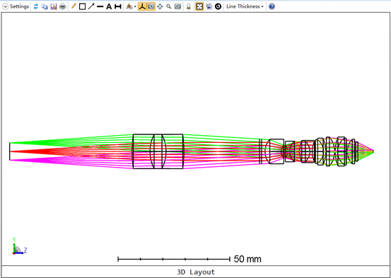
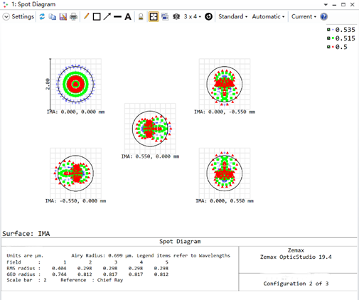
Fluorescence biological microscope objective aberration analysis diagram designed by Avantier
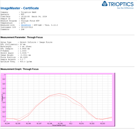
MTF test image of fluorescent biological microscope objective designed by Avantier
3. Specialized Coatings and Materials
Broadband Anti-Reflective Coatings: Multi-layer films (MgF₂, SiO₂) increase transmittance to >95% within the 350–800 nm range.
Low Autofluorescence Materials: Designers select specialized glass (e.g., fused silica or quartz) to prevent “self-illumination” of the lens, which would otherwise degrade the signal-to-noise ratio.
II. Core Functionalities and Performance
High-Resolution Detail Imaging
Through high NA and precise correction, these objectives reach the diffraction limit. According to the Rayleigh criterion (d = 0.61λ / NA), a 100x oil objective can resolve structures down to 0.2 μm, allowing for the visualization of mitochondria and ribosomes.
High-Efficiency Signal Acquisition
Weak fluorescence signals are a major bottleneck. Immersion designs (oil/water) minimize refractive index mismatches, allowing for the capture of single-molecule fluorescence. Water immersion, specifically, improves collection efficiency by over 30% compared to air lenses for deep-tissue imaging.
Low Phototoxicity and Sample Preservation
Live specimen imaging requires a delicate balance. High-efficiency light collection allows researchers to use lower excitation power, significantly reducing photobleaching and phototoxicity, thereby preserving the natural state of the cell.
Multi-Band and Multi-Mode Compatibility
Modern objectives are compatible with diverse imaging modalities:
Confocal Microscopy: Requires high transmission and pinhole accuracy.
TIRF (Total Internal Reflection Fluorescence): Requires high-angle incident light to excite only the sample surface (within ~100 nm).
FRET & Colocalization: Requires perfect chromatic registration across UV and NIR bands.
The 20X fluorescence microscope objective designed by Avantier clearly displays the 9 groups of No.6 on the resolution plate.
III. Primary Application Fields
Field
Application Focus
Key Requirement
Cell Biology
Organelle morphology, protein localization.
High resolution (NA > 1.3).
Neuroscience
Synaptic connections, calcium signaling.
High sensitivity & deep tissue penetration.
Developmental Biology
Embryonic cleavage, organogenesis.
Long working distance & low phototoxicity.
Clinical Diagnostics
Pathogen detection, tumor markers (HER2).
High contrast & field uniformity.
Microbiology
Viral invasion, bacterial colonization.
Super-resolution (STED/SIM) compatibility.
1. Neuroscience and Calcium Imaging
By utilizing fluorescent calcium indicators and high-sensitivity objectives, researchers can monitor real-time neuronal firing. This is essential for mapping neural networks and understanding signal transmission in the brain.
2. Clinical and Pathological Diagnosis
In immunofluorescence staining, objectives help identify specific antigens in biopsies. This is a gold standard for diagnosing malignancies and viral infections, providing rapid results with high specificity.
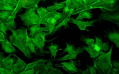
Fibroblast images observed through Avantier’s fluorescence biological microscope
IV. Conclusion and Future Outlook
The evolution of the fluorescence biological microscope objective—from basic glass elements to high-NA, apochromatic precision tools—has been the engine of discovery in life sciences. As we move toward super-resolution (nanoscale) and deep-tissue in vivo imaging, the next generation of objectives will focus on broader spectral ranges and further miniaturization for integrated “lab-on-a-chip” systems.