
High resolution and long working distance objective plays a crucial role in microscopy systems for medical device, scientific research, and industrial applications. Avantier Inc. is a leading manufacturer of producing high quality microscope objective lenses especially high NA objective lenses, long working distance objective lenses, large FOV objective lenses, and APO objective lenses.
To improve the imaging performance and reduce the optical aberrations, Avantier uses the active alignment method for meeting the high resolution requirements at the final assembling stage. Active alignment requires a special device with high imaging resolution.
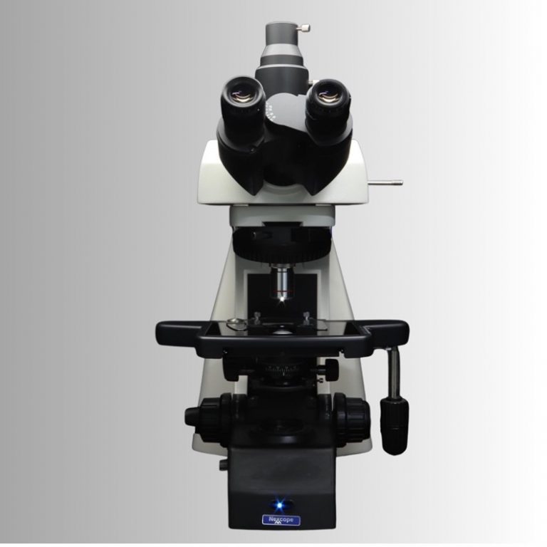
A setup for active alignment is shown in Figure 1. An infinite corrected objective lens with three screwed holes is assembled and mounted in a microscopy imaging system for testing. A star target with pinholes of 1 ~ 3 microns is placed on the stage and slightly defocused for the objective lens. A high resolution camera is on the top to capture images of the star target.
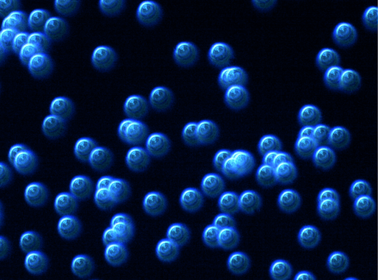
Figure 2 shows the captured star image before operating active alignment.
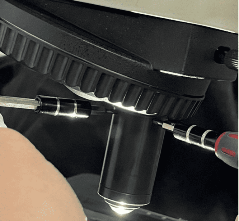
Figure 3 shows the objective lens with special alignment tool installed for aligning the most sensitive lens component to eliminating the lens aberration and improve the objective performance.
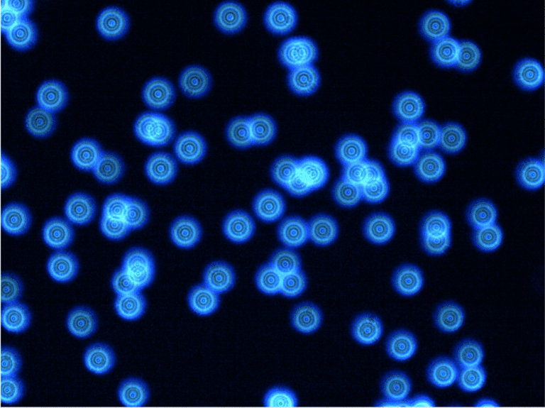
Figure 4 shows the star image after the active alignment method. The star image shows all the spots with round and concentric rings.

Figure 5 shows a typical image of the star target at the focus position.

Figure 6 shows a typical image of the objective lens with a resolutionabove 1000 lp/mm after active alignment.
Active alignment is a technology that improves the optical performance and yield in manufacturing advanced optical lenses. Avantier usually recommends active alignment for high resolution imaging lenses and high NA objective lenses.
Key Terms
High-resolution
In the realm of optical microscopy, high resolution is paramount. Microscopes equipped with high-resolution objectives are indispensable tools in fields such as biology, medicine, and materials science. APO objective lenses allow scientists to peer into the microscopic world with extraordinary clarity, revealing intricate cellular structures, microorganisms, and nanoparticles. High-resolution microscopy has been instrumental in advancing medical research, enabling the diagnosis of diseases at earlier stages and facilitating breakthroughs in drug development.
High-resolution optical systems are also indispensable in various industries, such as semiconductor manufacturing and lithography. These systems use advanced optics to create intricate patterns on microchips with nanometer-level precision. The ability to achieve high resolution is critical for producing smaller and more powerful electronic devices, driving innovation in technology.
Furthermore, high-resolution optics are central to the field of spectroscopy, where they enable scientists to analyze the properties of materials and molecules at the atomic and molecular levels. High-resolution spectroscopy plays a vital role in chemistry, environmental science, and materials science, aiding in the identification of compounds, the study of chemical reactions, and the development of new materials.
Long Working Distance Objectives
Long working distance objectives are a fundamental component in microscopy that play a pivotal role in a wide range of scientific and industrial applications. These specialized optical lenses provide a significant advantage by allowing microscope to examine samples with ample space between the objective lens and the specimen. In this article, we will explore the importance of long working distance objectives and their applications in microscopy.
Long working distance objectives are characterized by their ability to maintain focus and capture clear images even when there is a substantial gap between the front lens element and the sample. This generous working distance, typically ranging from a few millimeters to several centimeters, provides numerous benefits in various microscopy scenarios.
One of the primary advantages of long working distance objectives is their versatility in accommodating diverse sample types and experimental setups. In life sciences, where researchers often work with living organisms or delicate specimens, these objectives enable the manipulation of samples without physically touching them. This minimizes the risk of contamination and damage, making long working distance objectives invaluable in fields like cell biology and microbiology.
In materials science and industrial applications, long working distance objectives facilitate the examination of three-dimensional objects, such as mechanical components or semiconductor wafers. The ample space between the lens and the sample allows for the use of specialized equipment, such as manipulators or measurement tools, which can be positioned precisely to interact with the specimen while under observation.
Another significant application of long working distance objectives is in surface metrology and profilometry. These objectives enable precise measurement of surface roughness, topography, and height variations on a wide range of materials. Researchers and engineers can use them to assess the quality of manufactured products, ensuring compliance with specifications and quality control standards.
High Numerical Aperture (NA)
High Numerical Aperture (NA) objective lenses are marvels of optical engineering, empowering scientists and researchers to delve deeper into the microscopic world with unprecedented precision and clarity. These specialized lenses have a profound impact across a spectrum of scientific disciplines, pushing the boundaries of what we can observe and understand. In this article, we will explore further the significance and applications of high NA objective lenses.
The Essence of Numerical Aperture (NA)
Numerical Aperture is a fundamental parameter in microscopy, representing the lens’s ability to collect and focus light from a specimen onto the image plane. It is a critical factor in determining the resolution and quality of microscopic images. High NA objective lenses are characterized by their ability to gather light from wide angles, enabling them to capture fine details and achieve superior image sharpness.
- Advancing Resolution and Image Quality – High NA objective lenses are revered for their capacity to deliver exceptional resolution. They excel at capturing intricate structures and minute features, making them indispensable in disciplines where precision matters. Biologists can explore cellular organelles and subcellular structures with greater detail, while materials scientists can characterize nanomaterials at the atomic level.
- Fluorescence Microscopy Elevated – In fluorescence microscopy, high NA objectives shine as stars. They enhance the brightness and resolution of fluorescent signals, facilitating the detection of specific molecules and structures within biological specimens. This capability is pivotal in life sciences, allowing researchers to study intricate cellular processes, protein interactions, and gene expression patterns.
- Confocal Microscopy’s Best Companions – Confocal microscopy, renowned for its ability to create three-dimensional images with optical sections, relies heavily on high NA objectives. These lenses enable researchers to capture thin optical slices with remarkable clarity, which is crucial for reconstructing detailed 3D structures. Applications span neuroscience, cell biology, and materials science, where in-depth visualization is vital.
- TIRF Microscopy Illuminated – Total Internal Reflection Fluorescence (TIRF) microscopy harnesses the power of high NA objectives to perfection. By creating a thin illumination layer at the specimen surface, TIRF microscopy excels at studying cellular processes at interfaces, such as the cell membrane. Researchers use it to unravel the mysteries of membrane dynamics, single-molecule behavior, and molecular interactions.
- Materials Science and Nanotechnology Insights – High NA objectives are pivotal in materials science and nanotechnology. They empower researchers to examine nanoscale materials and structures with unparalleled precision. The analysis of nanoparticles, nanowires, and thin films benefits immensely from these objectives, aiding in the development of advanced materials and devices.
Large Field of View (FOV) Objective Lenses
Large Field of View (FOV) objective lenses are instrumental components in microscopy, offering researchers a broader perspective and deeper insights into samples, be they biological specimens or materials. These specialized optics provide a panoramic view, enabling scientists to visualize more extensive areas of interest without sacrificing image quality. In this article, we’ll delve into the significance and applications of large FOV objective lenses.
- Unveiling Field of View (FOV) – Field of View (FOV) in microscopy refers to the area of the specimen that can be observed through the microscope’s objective lens at any given magnification. It is a critical parameter that directly impacts the extent of the sample that can be examined in a single view. Large FOV objective lenses are designed to expand this viewing area significantly.
- A Wider Perspective – One of the primary advantages of large FOV objective lenses is their ability to provide a more comprehensive view of samples. Researchers can observe larger portions of specimens, which is particularly advantageous when studying tissues, geological samples, or materials with extensive features. This broader perspective allows for improved context and a deeper understanding of the sample’s structure and characteristics.
- Efficiency in Scanning and Mapping – Large FOV objectives are valuable in various scientific disciplines, including geology and materials science. Geologists can use these lenses to survey rock formations and minerals more efficiently, while materials scientists can map the properties and defects in materials with a broader scope. In manufacturing and quality control, large FOV objectives aid in inspecting larger sections of products, facilitating faster and more comprehensive assessments.
- Microscopy Imaging in Industrial Settings – Large FOV objectives find applications in industrial microscopy, where quality control and defect analysis are paramount. These objectives allow for the inspection of more extensive surfaces, aiding in the detection of defects, imperfections, or deviations from desired specifications in manufactured components and materials.
APO Objective Lenses
- Precise Color Reproduction – One of the most remarkable features of APO objective lenses is their ability to deliver accurate and true-to-life color reproduction. This is especially critical in fields like biology and materials science, where precise color information can be crucial for identifying cellular structures, staining, or materials properties. APO lenses ensure that colors in the sample are faithfully and consistently represented in the images, eliminating the color distortions associated with other types of objectives.
- Sharper and More Detailed Images – APO lenses are also renowned for their exceptional resolution. By correcting chromatic aberration as well as other optical imperfections like spherical and comatic aberrations, these lenses provide sharper, higher-contrast images with finer details. This level of clarity is vital in applications such as histology, where distinguishing subtle cellular structures is essential.
- Fluorescence Imaging – In fluorescence microscopy, APO objective lenses are indispensable. They allow researchers to capture the full spectral range of fluorescent dyes without introducing color artifacts. This capability is crucial for multicolor fluorescence imaging experiments, where precise color separation is essential for identifying different fluorophores and studying cellular processes with high specificity.
- Confocal and Super-Resolution Microscopy – APO objectives are commonly used in advanced microscopy techniques like confocal and super-resolution microscopy. These techniques demand exceptional image quality and precision to achieve their objectives. APO lenses play a pivotal role in minimizing optical distortions and delivering the sharpness required for three-dimensional imaging and the visualization of nanoscale structures.


GREAT ARTICLE!
Share this article to gain insights from your connections!